The cornea is a structure formed by two optical surfaces. Correct corneal morphology allows visual transparency and provides corneal dioptric power. However, in some corneal pathologies or ectasias such as keratoconus, the corneal integrity is compromised. In such situations, to slow the progression of keratoconus various treatments as contact lenses, cross-linking or intrastromal rings among others have been developed. If it is not possible to slow down the progression of keratoconus by means of the previous treatments, keratoplasty surgery will be necessary. Nevertheless, keratoconus is not the only corneal ectasia or pathology where a corneal transplant is necessary. Due to the importance of achieving good visual outcomes after corneal transplant, different procedures as Penetrating Keratoplasty (PK), Deep Anterior Lamellar Keratoplasty (DALK) or Posterior Lamellar Keratoplasty (PLK) have been developed. Likewise, the improvements made in the keratoplasty technology have allowed to achieve better visual outcomes and to reduce the postoperative corneal complications. For example, the introduction of the femtosecond laser in keratoplasty surgery stands out by its safety and precision. Precisely, in module 3 of corneal surgery of the Online Course entitled Clinical Methodology in Refractive Cataract and Corneal Surgery, the different techniques and important innovations of corneal keratoplasty are widely explained.
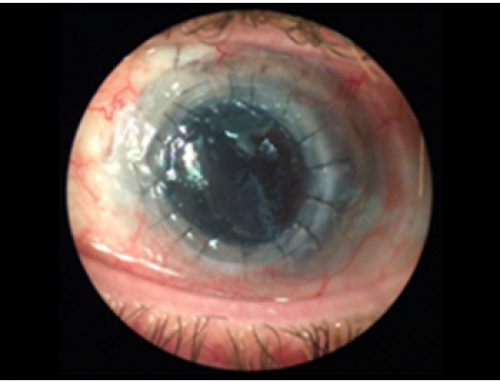
What devices are useful to evaluate the corneal integrity in keratoplasty surgery?
To evaluate the possibility of corneal keratoplasty to recover the corneal integrity, in addition assessing visual acuity, other diagnostic tests are required. Concretely, corneal topography is very useful to evaluate the corneal aberrometry and power. Devices that analyse both corneal surfaces like Sirius or Pentacam are very useful because provides more information about corneal integrity. However, corneal topography is not only useful in the preoperative of corneal keratoplasty, but also postoperatively. Likewise, Optical Coherence Tomography (OCT) is another diagnostic device very useful in ophthalmologic consultation to evaluate the integrity and position of the corneal graft postoperatively. Due to the importance of interpreting the different diagnostic images obtained in ophthalmologic consultation, an appropriate training in keratoplasty surgery is necessary. Concretely, in the Online Course entitled Clinical Methodology in Refractive Cataract and Corneal Surgery, we provide an adequate and complete training program on keratoplasty surgery. Our international tutors are highly experienced professionals in clinical practice, research, and teaching. Professor Jorge L. Alió, have performed hundreds of keratoplasty surgeries in Spain and internationally and has published more than 500 papers/ peer review scientific articles in Pubmed Central. Likewise, the other international tutors have participated in important international congresses on keratoplasty surgery, and have also published some scientific articles about this topic. For these reasons, our international course recognized with 25 ECTS credits, covers all the needs for the proper training of the ophthalmologists in keratoplasty surgery.
Some scientific references of the course about this topic
Alio JL, Abdelghany AA, Abu-Mustafa SK, Zein G. A new epidescemetic keratoprosthesis: pilot investigation and proof of concept of a new alternative solution for corneal blindness. Br J Ophthalmol. 2015 Nov; 99(11):1483-7.
Alio JL, Abdelghany AA, Barraquer R, Hammouda LM, Sabry AM. Femtosecond Laser Assisted Deep Anterior Lamellar Keratoplasty Outcomes and Healing Patterns Compared to Manual Technique. Biomed Res Int. 2015; 2015:397891.
Arnalich-Montiel F, Alió Del Barrio JL, Alió JL. Corneal surgery in keratoconus: which type, which technique, which outcomes? Eye Vis (Lond). 2016 Jan 18;3:2.