Keratoconus is a corneal disease characterized by progressive thinning corneal stroma, which produces increased corneal power and corneal aberrations. Different treatments have been developed to reduce the progression of keratoconus and delay keratoplasty, as intrastromal corneal rings or crosslinking among others. Advances in crosslinking procedure, as likewise in clinical devices facilitate the monitoring of the pathology, and require be updated in clinical practice.
What progress has been made in crosslinking procedure?
Crosslinking is a photochemical treatment that reduces the progression of corneal ectasia, especially recommended in progressive keratoconus. The advent of femtosecond laser allows among its many functions the implantation of intrastromal rings for the creation of the tunnel, as the realization of the pockets in cross-linking. Indeed, the new femtosecond lasers allow that the realization of the pocket in crosslinking procedure is visualized by the integrated Optical Coherence Tomography (OCT).
By installing riboflavin illuminated with ultraviolet light for about 30 minutes it allows the collagen fibers are strengthened. Two types of crosslinking procedure have been developed, standard and accelerated, both showing increased corneal enzymatic resistance. Precisely in a recent article published in Acta Ophthalmology, Dr David O’Brart proved that the modification to the standard iontophoresis trans-epithelial technique provided successful penetration of riboflavin into the stroma. Furthermore, in the same study published by the professor of the “Online Course Clinical Methodology in Refractive Cataract and Corneal Surgery” determined that it appears to offer the most promise for epithelium-on crosslinking.

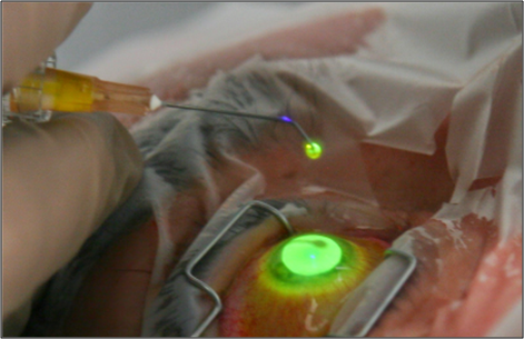
Top image: Iontophoresis. Bottom image: Application of riboflavin in a patient with keratoconus.
Scientific studies outcomes show that the combination of crosslinking with implantation of intrastromal rings in the same intervention provides better results, reduces the progression of keratoconus, and delays the realization of keratoplasty.
How to improve the technology of new diagnostic devices in keratoconus patients operated by cross-linking?
Corneal topographers are suitable to evaluate the effect of crosslinking for the treatment of keratoconus, especially those that analyze both corneal surfaces. By corneal topography the examiner can analyze the variations of corneal thickness, as likewise the corneal diopter power in keratoconus patients operated by crosslinking. In addition, those corneal topographers that incorporate analysis of aberrations and optical quality allow analyzing and documenting the variation in time. Aberrometers systems also provide useful information of optical quality (Point Spread Function, Modulation Transfer Function…), as corneal and ocular aberrations in keratoconus patients operated by crosslinking. Specifically, crosslinking in keratoconus patients improvement topographic and wavefront parameters and even are maintained at 7 years according one study published by Dr David O’Brart in the American Journal of Ophthalmology.
In addition to the aforementioned devices, confocal microscopy and Anterior Segment Optical Coherence Tomography (AS-OCT) have shown high value in the assessment of corneal and ocular parameters after crosslinking treatment in keratoconus patients. Indeed, AS-OCT is also special diagnostic value because has been found that after crosslinking treatment the depth of the anterior chamber is reduced. Due to this appreciation, will be necessary delay phakic intraocular lens implantation if is necessary correct the refractive error typical in keratoconus patients.
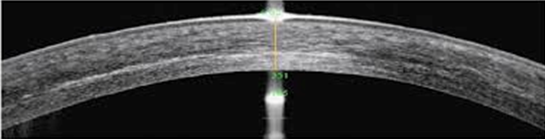
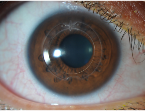
Crosslinking demarcation line performed in a patient with keratoconus.
In the Online Course Clinical Methodology in Refractive Cataract and Corneal Surgery through the extensive educational material provides high-quality training on the surgical procedure of cross-linking in keratoconus patients, as well pharmacological treatment and therapy after surgery. The course is integrated by more than 40 international tutors of high clinical and scientific prestige, which provide exceptional rigor to the training program. In addition, the entire program is online, it can be studied from any country in the world, and it is recognized by 25 ECTS credits by University Miguel Hernandez.
Bibliographic references
Aldahlawi NH, Hayes S, O’Brart DP, Meek KM. Standard versus accelerated riboflavin-ultraviolet corneal collagen crosslinking: Resistance against enzymatic digestion. J Cataract Refract Surg. 2015 Sep;41(9):1989-96.
Hayes S, Morgan SR, O’Brart DP, O’Brart N, Meek KM. A study of stromal riboflavin absorption in ex vivo porcine corneas using new and existing delivery protocols for corneal cross-linking. Acta Ophthalmol. 2016 Mar;94(2):e109-17.
O’Brart DP, Kwong TQ, Patel P, McDonald RJ, O’Brart NA. Long-term follow-up of riboflavin/ultraviolet A (370 nm) corneal collagen cross-linking to halt the progression of keratoconus. Br J Ophthalmol. 2013 Apr;97(4):433-7.
O’Brart DP, Patel P, Lascaratos G, Wagh VK, Tam C, Lee J, O’Brart NA. Corneal Cross-linking to Halt the Progression of Keratoconus and Corneal Ectasia: Seven-Year Follow-up. Am J Ophthalmol. 2015 Dec;160(6):1154-63.