In patients with corneal ecstasies as keratoconus, one of the alternatives to regularize the corneal surface is the implantation of intrastromal corneal rings. As it is known keratoconus produce an increase of the corneal curvature and a decrease in corneal thickness, among other alterations. According to keratoconus evolution grade, intraestromal corneal ring features will be different in thickness, length and diameter. To choose the parameters of intrastromal corneal rings, advances in diagnostic devices provide important information about the characteristics of the cornea preoperatively. For example, some corneal topographers with Scheimpflug imaging in addition to providing topographic and aberrometric parameters, include intrastromal corneal ring module which is highly useful. However, there are other devices highly useful in the diagnosis and monitoring of patients with keratoconus who are implanted intrastromal corneal rings. Devices that analyze ocular biomechanics have been consolidated mainly for the evaluation in keratoconus patients in clinical practice and research because it allows evaluating in the time the ocular response. Other devices such as endothelial microscopy, confocal microscopy, ocular aberrometry, or optical coherence tomography among others provide information very useful in patients with keratoconus and intrastromal corneal rings, which be explained in detail in “Online Course Clinical Methodology in Refractive Cataract and Corneal Surgery” by experimented international tutors.
Top image: Triangular profile of Ferrara rings (Kerarings). Bottom image: Hexagonal profile of Intacs.
What are the differences between intrastromal corneal rings?
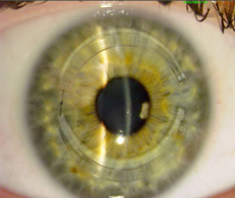
Different intrastromal corneal rings that allows regulate the progression of corneal ectasia mainly keratoconus, which differ in their characteristics and parameters have been developed. An example of intraestromal corneal ring is VissumRing (VR- technology) shown in the superior image of this publication, innovation developed by our Department of Investigation of VISSUM Instituto Oftalmológico de Alicante in conjunction with Mediphacos. Characteristics of the intrastromal corneal rings vary depending on the refractive errors or spherical equivalents according to manufacturers nomograms. In all intrastromal corneal rings, by inserting it seeks flatten the corneal surface in order to reduce corneal power. However, the effect produced depends on the thickness of the ring, its diameter, and its centering mainly.
Top image: Intacs. Bottom image: Myoring technology.
All differences between intrastromal corneal rings as presurgical plan for insertion are duly explained in “Online Course Clinical Methodology in Refractive Cataract and Corneal Surgery”.
What improvements have been developed in the surgery of intrastromal rings?
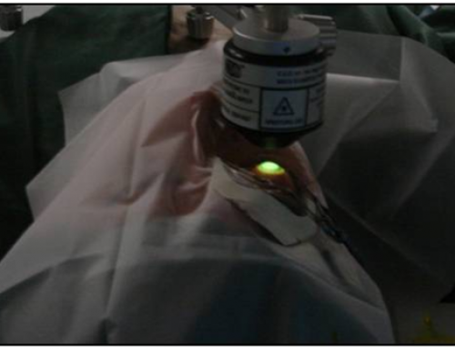
In the surgical procedure of implantation of intrastromal corneal rings significant improvements in the technology have been made. The use of femtosecond laser to perform the incision and tunnels where are introduced intrastromal corneal rings, providing increased safety, effectiveness and accuracy. Currently, in the intervention of intrastromal corneal rings, also crosslinking can be performed to stabilize the ectasia. Even later it can be possible performed topography-guided transepithelial PRK, or implant phakic Intraocular Lenses (IOLs) to correct the residual refractive error. One of our international tutors of the course, Dr George Kymionis, published an article in Journal Cataract & Refractive Surgery, in what proved that toric implantable collagen copolymer phakic IOLs after crosslinking and corneal ring segment in patients with keratoconus, was effective and improved functional vision reducing the progression of the ectasia. Besides the above, intrastromal corneal rings have the advantage that they can be replaced if the effect achieved is not desired or complications occurred. Indeed, another of our prestigious international tutors of the course, Dr Mahmoud Ismail participated on 19-21 of January 2016 in a presentation in the Research Institute of Ophthalmology of the Cairo about Myoring implantation following failed intracorneal ring segments.
During the “Online Course Clinical Methodology in Refractive Cataract and Corneal Surgery”, our prestigious and experimented international tutors will provide enough information about the different diagnostic devices needed for pre- and post-surgical evaluation in patients with keratoconus with intraestromal corneal rings. Also the different improvements made in the surgical procedure of implantation of intraestromal corneal rings are especially evaluated, as their results, and possible complications are discussed.
Bibliographic references
Coskunseven E, Jankov MR 2nd, Grentzelos MA, Plaka AD, Limnopoulou AN, Kymionis GD. Topography-guided transepithelial PRK after intracorneal ring segments implantation and corneal collagen CXL in a three-step procedure for keratoconus. J Refract Surg. 2013 Jan;29(1):54-8.
Coşkunseven E, Sharma DP, Jankov MR 2nd, Kymionis GD, Richoz O, Hafezi F. Collagen copolymer toric phakic intraocular lens for residual myopic astigmatism after intrastromal corneal ring segment implantation and corneal collagen crosslinking in a 3-stage procedure for keratoconus. J Cataract Refract Surg. 2013 May;39(5):722-9.
Diakonis VF, Kankariya VP, Woreta F, Yoo SH, Lubahn JG, Kymionis GD et al. Refractive and topographic fluctuations due to intracorneal ring segments motility. J Refract Surg. 2014 Feb;30(2):140-2.