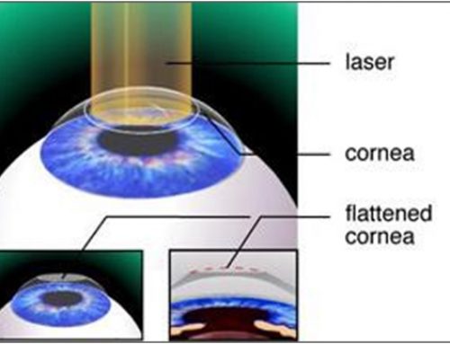
Laser-Assisted In Situ Keratomileusis (LASIK) is one of the surgical ambulatory procedures most performed worldwide for the correction of myopia, hyperopia or astigmatism. In the surgery it is necessary to lift a corneal lenticule with a femtosecond laser for then apply excimer laser technology on the patient’s corneal stroma. Nevertheless, not all the patients are candidates to perform the surgery due to the ablation depth depend on the diopters, as the thickness, aberrometry, and morphology of the cornea. Concretely, to know if a patient is suitable for surgery, corneal topography highlights among the diagnostic necessary devices in a consultation of refractive surgery. Specifically, topographers which analyze both corneal surfaces are particularly useful in LASIK refractive surgery. Likewise, it is also necessary to perform a bimicroscopic examination by means of slit-lamp to evaluate the cornea and logically graduate patient’s vision.
However, the success of the procedure in the quality of vision of the patient depends on the diagnostic and surgical technology employee. Since the introduction of LASIK in 1990 different improvements realized in the technology have allowed increase notably the success rate, satisfaction, efficacy and safety of the procedure. Indeed, Prof. Dr. Jorge Alió, Director of Online Course Clinical Methodology in Refractive Cataract and Corneal Surgery has one of the largets experiences in Spain both in the clinical practice and in the clinical research of the LASIK procedure. In fact, he was one of the worldwide pioneers in LASK and he was the one who installed the first Excimer Laser 20/20 B installing the VISX 20/20 B excimer laser platform in Spain in 1992. Since then, he and his group in Vissum Alicante have been constantly updated on the technique and proposing innovations to make LASIK safer and more precise .
What advances have been developed in the LASIK surgical procedure?
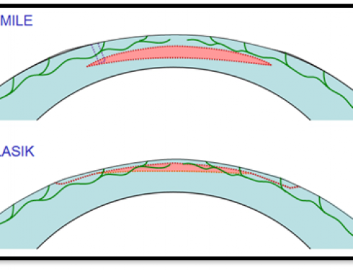
Since the beginning of the procedure, LASIK has experienced significant advances, as for example the advent of femtosecond laser for performing corneal flap. Among the multiple advantages provided by the femtosecond laser in corneal refractive surgery highlights lower incidence of dry eye, reduced complications, as faster recovery of visual quality. Precisely, the visual recovery after the application of the femtosecond laser is significantly higher compared when microkeratome is used to perform the corneal lenticule. Realized with the microkeratome or with femtosecond laser the corneal flap, proceed to the application of excimer laser. There are also notable advances that the technology of the excimer laser has experienced in the procedure of LASIK refractive corneal surgery. Stands out mainly the introduction of platforms that provide faster speed of corneal ablation, shorter duration of the procedure, better eye-tracker systems, or higher diopter correction, among others… Concretely, in a recent scientific study published in the Journal Refractive Surgery by the international tutor of the UMH course, Dr. Dan Reinstein proved that MEL 80 excimer laser provided high efficacy and safety for the treatment of high myopia among -8.00and -14.25 diopters and up to -4.50 diopters of cylinder. The findings reported by Dr. Reinstein have further confirmed the reports published by Prof. Jorge Alio and Dr. Alfredo Vega about the good outcomes of Lasik in adequately selected myopes. Among other advantages, the advances developed in the modern refractive surgery lasers allow better visual quality after the procedure, reduce spherical aberration, as likewise provide better night and contrast vision. Finished the application of the excimer laser for the correction of the refractive errors, the corneal flap is repositioned, and some postoperative reviews will be necessary to know the visual quality obtained after the procedure.
Due to the advances developed in the LASIK for the correction of refractive errors, it is necessary to be continuously formed in the procedure as well in the managing of the different diagnostic devices. Concretely the high clinical experience, the research background, and experience in teaching of the international tutors of the first online course about refractive and cataract surgery guarantees the quality of the course contents.
Bibliographic references
Alió JL, Muftuoglu O, Ortiz D, Pérez-Santonja JJ, Artola A, Ayala MJ et al. Ten year follow-up of laser in situ keratomileusis for myopia of up to -10 diopters. Am J Ophthalmol. 2008 Jan;145(1):46-54.
Alio JL, Vega-Estrada A, Piñero DP. Laser-assisted in situ keratomileusis in high levels of myopia with the amaris excimer laser using optimized aspherical profiles. Am J Ophthalmol. 2011 Dec;152(6):954-963.e1.
Reinstein DZ, Carp GI, Archer TJ, Gobbe M. Transitioning from mechanical microkeratome to femtosecond laser flap creation: visual outcomes of an experienced and a novice LASIK surgeon. J Cataract Refract Surg. 2012 Oct;38(10):1788-95.
Reinstein DZ, Carp GI, Archer TJ, Lewis TA, Gobbe M, Moore J et al. Long-term Visual and Refractive Outcomes After LASIK for High Myopia and Astigmatism From -8.00 to -14.25 D. J Refract Surg. 2016 May 1;32(5):290-7.
Reinstein DZ, Carp GI, Lewis TA, Archer TJ, Gobbe M. Outcomes for Myopic LASIK With the MEL 90 excimer laser. J Refract Surg. 2015 May;31(5):316-21.